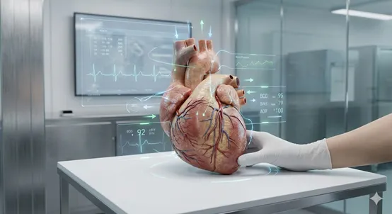
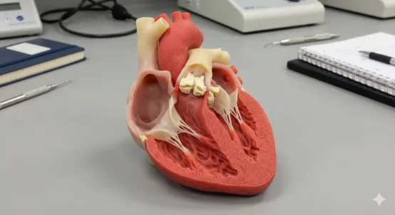
An anatomical 3D model is a physical or digital representation of an organ, system, or anatomical structure of the human body. These models are used across medicine and medical technology for visualization, education, training, research, and clinical decision-making.
Some models are generic, based on standard human anatomy. Others are patient-specific, created directly from medical imaging data such as CT or MRI scans. That distinction, generic versus personalized, is where these models shift from teaching tools into decision-making instruments.
A short historical perspective
For centuries, anatomy has been taught through physical models. Early examples include wax sculptures, bone replicas, and hand-crafted models made from wood, plaster, or clay. During the 20th century, industrially produced plastic models became standard equipment in medical schools worldwide.
The real transformation arrived in the 21st century. Advanced imaging technologies, CT, MRI, and ultrasound, combined with digital segmentation and 3D printing made it possible to create highly accurate, patient-specific anatomical models. For the first time, clinicians could hold a real-scale representation of a specific patient's anatomy in their hands.
Medicine moved from "average anatomy" to individual anatomy.
How anatomical 3D models are used today
Medical education and clinical training
Anatomical models give students, residents, and clinical teams a clear spatial understanding of complex structures.
Value: three-dimensional comprehension is significantly stronger than learning from 2D images alone.
Pre-operative surgical planning
Patient-specific models let surgeons rehearse complex procedures, test surgical approaches, and evaluate implant positioning before entering the operating room.
Value: better preparation, reduced uncertainty, and improved team communication.
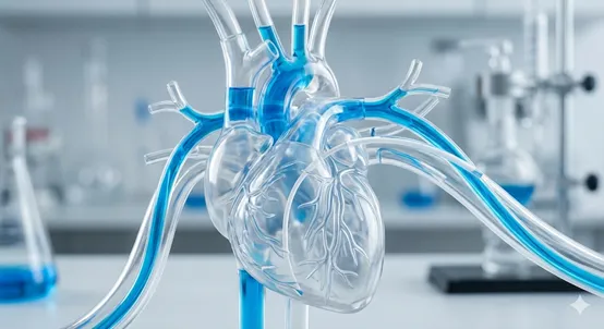
Medical device R&D and engineering validation
Medical device companies use anatomical models to simulate how tissues interact with implants, catheters, or delivery systems.
Value: faster iteration, earlier detection of design issues, and safer development cycles.
Patient communication
Anatomical models help explain conditions and procedures to patients and their families in a clear, visual way.
Value: informed consent and stronger trust.
Product demonstration and investor communication
For medical technology companies, physical models turn complex engineering concepts into something tangible.
Value: clarity, credibility, and impact.
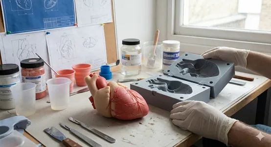
From medical imaging to physical reality
Creating an anatomical 3D model usually starts with medical imaging data.
Digital modeling and segmentation
CT or MRI scans are processed through segmentation, a digital process that isolates specific anatomical structures from imaging data. Accuracy at this stage defines everything that follows.
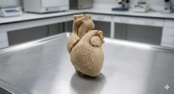
3D printing
Advanced 3D printing enables the production of:
- rigid, bone-like structures
- flexible vessels
- transparent or multi-material models
Physical interaction reveals insights that screens often miss.
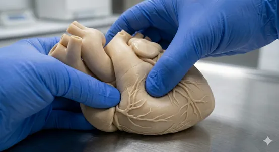
Silicone casting and soft-tissue simulation
Silicone and polymer casting techniques create models that mimic the feel and behavior of real tissue, essential for procedural training and device testing under realistic conditions. See the material range.
Virtual and augmented reality (VR / AR)
Interactive digital models can be explored in immersive environments for planning, training, and collaboration, enabling remote access and repeatable simulations without physical wear.
Generic vs. patient-specific anatomical models
Generic models
Standardized models based on average human anatomy, typically mass-produced.
- Low cost
- High availability
- Reusable
- Do not reflect anatomical variability
- Cannot represent unique pathologies
- Education and general demonstrations
Patient-specific models
Derived from a single patient's CT, MRI, or ultrasound data.
- Reflect real anatomy, including deformities or rare conditions
- Enable precise planning and testing
- Higher cost
- Depend on imaging quality and expert processing
- Complex surgery, personalized medicine, device development
Challenges and considerations
- Cost: specialized software, equipment, and expertise are required.
- Technical limits: perfect replication of biological tissue properties is still evolving.
- Data ethics: patient privacy and data protection are critical.
Understanding these constraints is essential for responsible and effective use.
Why anatomical 3D models matter
Generic models help us learn anatomy. Patient-specific models help us make better decisions. They bridge the gap between medical imaging and real-world action, whether that action happens in the operating room, the R&D lab, or a design-review meeting.